
The Sleeping Brain: A Window Through the Pupil
For years, sleep has been considered a period of rest and recuperation, a time when our bodies and minds power down to recharge. However, recent research challenges this simplistic view, revealing that our brains remain surprisingly active during sleep. A team of researchers in Switzerland has discovered that eye movements, specifically pupil size variations, during sleep offer a fascinating window into the dynamic processes occurring within the sleeping brain. Their study, published in Nature Communications, sheds light on the intricate relationship between pupil dynamics and brain activity, potentially paving the way for new diagnostic tools for sleep disorders and other neurological conditions.
The conventional understanding of sleep assumes a state of reduced brain activity. But Caroline Lustenberger, a neuroscientist at ETH Zürich and co-author of the study, points out that their findings contradict this assumption. According to the research, the pupils of sleeping individuals constantly change size, indicating fluctuating levels of brain activation. These fluctuations are the brain’s response to various stimuli, suggesting that the sleeping brain is far from inactive. Instead, it is constantly processing information and responding to its internal and external environment.
The significance of pupil dynamics lies in their connection to the locus coeruleus, a brain region located in the brainstem that plays a crucial role in regulating arousal levels. This region is notoriously difficult to study directly in sleeping individuals. However, researchers have long known that pupil size is an indicator of brain activity. Lustenberger and her colleagues hypothesized that they could use pupil dynamics to indirectly track brain activation during sleep, offering a novel approach to understanding the complex processes that unfold during our slumber.
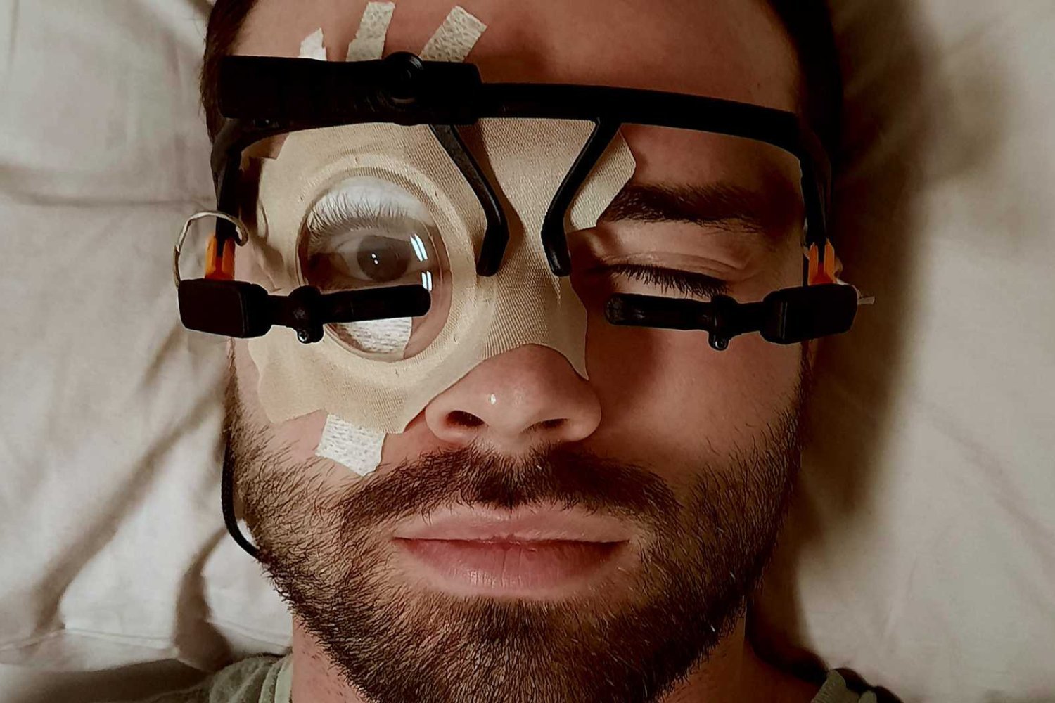
To conduct their study, the researchers developed a unique technique to observe eye movements for extended periods during sleep. The innovative methodology involved taping one eye of each participant open, moistening it with eye ointment, and sealing it behind a transparent bandage. This allowed them to monitor pupil size variations continuously without disrupting the participants’ sleep.
Manuel Carro Domínguez, a biomedical engineer at ETH Zürich and lead author of the study, explained that the team was initially concerned that participants would be unable to sleep with their eyes open. However, they found that in a dark room, most people quickly forgot about their open eye and were able to fall asleep comfortably. This ingenious technique enabled the researchers to gather valuable data on pupil dynamics during sleep.
The results of the study revealed that pupil size constantly changes throughout the sleep cycle, indicating that brain activation levels are not constant but rather fluctuate continuously. This finding confirms a biological feature previously observed in rodents, suggesting that this dynamic brain activity during sleep is a conserved mechanism across species.
Furthermore, the researchers identified a link between pupil dynamics during sleep and specific brain activity patterns. They found that pupil size variations were correlated with brain waves associated with sleep stability and memory consolidation. This suggests that pupil dynamics may reflect the brain’s efforts to maintain sleep and process information during the night.
The study also explored the brain’s response to external stimuli during sleep. The researchers discovered that the intensity of the brain’s reaction to sound depended on the level of brain activation, as indicated by the participants’ pupils. This suggests that the brain is not completely isolated from the outside world during sleep but rather continues to monitor its surroundings and respond to relevant stimuli.
While the study provided valuable insights into the relationship between pupil dynamics and brain activity, it did not establish a direct causal link between the locus coeruleus and pupil size variations. As Lustenberger clarified, the researchers are "simply observing pupil changes that are related to the level of brain activation and heart activity."
Despite this limitation, the researchers believe that their findings have significant implications for understanding sleep and its role in brain function. They plan to conduct a follow-up study to investigate the potential causal relationship between the locus coeruleus and pupil dynamics, as well as to explore how activation levels influence sleep quality and other cognitive processes.
If future research confirms a strong causal relationship, pupil movement could potentially be used as a diagnostic tool for a range of disorders, including insomnia, post-traumatic stress disorder, and even to assess the recovery of comatose patients. By monitoring pupil dynamics during sleep, doctors may be able to gain valuable insights into brain function and identify abnormalities that could indicate underlying medical conditions.
The study’s findings have opened up a new avenue for exploring the complexities of the sleeping brain. By focusing on the humble pupil, the researchers have provided a window into the dynamic processes that occur during sleep, challenging conventional assumptions and paving the way for new diagnostic and therapeutic interventions.
The eye, often described as the window to the soul, may also be a window to the brain. As this research demonstrates, the seemingly simple act of observing pupil size variations during sleep can reveal a wealth of information about the brain’s activity and its intricate relationship with the body.