
Here’s a longer, more detailed version of the article, formatted in Markdown and exceeding 600 words:
Challenging Dogma: New Research Suggests Cell Division Isn’t Always Symmetrical
For generations, the image of cell division, or mitosis, etched in the minds of high school biology students has been remarkably consistent. The textbook narrative depicts a parent cell transforming into a perfect sphere before elegantly splitting into two identical daughter cells, mirror images in size, shape, and function. This seemingly immutable process has served as a cornerstone of biological understanding for over a century. However, groundbreaking research published in the prestigious journal Science is poised to disrupt this long-held belief, potentially rewriting biology textbooks and reshaping our comprehension of fundamental cellular mechanisms.
The study, spearheaded by researchers at the University of Manchester, reveals that mitosis is not always characterized by cell rounding – the transformation of a parent cell into a spherical shape. This crucial deviation from the textbook model has profound implications, suggesting that daughter cells resulting from division are not invariably symmetrical or equipped with identical functions. The ramifications of this discovery extend far beyond the classroom, offering new insights into diseases such as cancer and the intricate processes of tissue and organ development.
Shane Herbert, co-lead author of the study and a researcher at the University of Manchester’s faculty of Biology, Medicine and Health, emphasized the discrepancy between textbook representations and the reality of cell division in living organisms. “Students learn that when a cell divides, it will generate a uniform spherical shape. Our study, however, shows that in real living organisms, it is not as simple as that,” Herbert stated. This revelation necessitates a re-evaluation of established models and opens up new avenues for exploring the complexities of cell behavior.
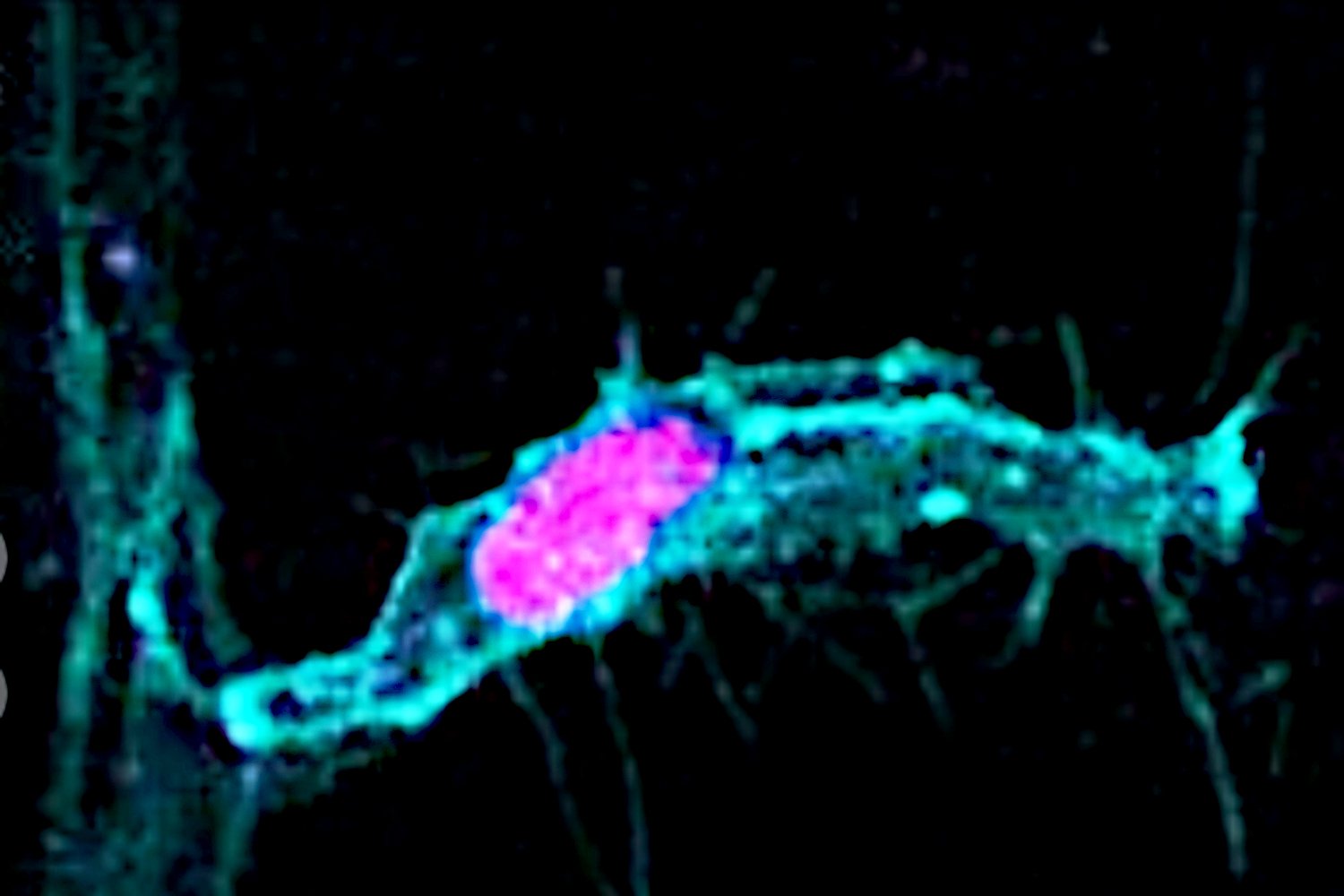
The researchers focused their investigations on blood vessel formation in zebrafish embryos, an ideal model system due to the transparency of the embryos, allowing for direct observation of cellular processes in vivo. The growth of new blood vessels involves the coordinated movement of cells, with slower-moving cells typically following a single, faster-moving "lead" cell. Intriguingly, when the lead cell underwent mitosis, it defied the conventional expectation of rounding. Instead, it divided asymmetrically, giving rise to two distinct daughter cells: one retaining the slower-moving characteristics of its predecessors and the other assuming the role of a new, fast-moving lead cell. This asymmetric division challenges the notion that mitosis invariably produces identical daughter cells and underscores the potential for functional diversity arising from cell division.
Holly Lovegrove, co-lead author of the study and a lecturer in cardiovascular sciences at The University of Manchester, highlighted the advantages of using zebrafish embryos for this type of research. “Using transparent 1-day old zebrafish embryos allows us to study a dynamic process like cell division inside a living organism,” Lovegrove explained. “We are therefore able to make movies of this fundamental cell behaviour and in doing so reveal exciting new aspects of how tissues grow.” The ability to visualize cell division in real-time within a living organism provides invaluable insights that would be difficult or impossible to obtain through other methods.
Further analysis by the researchers revealed a correlation between the shape of the parent cell and the symmetry of its division. Shorter, wider cells were more likely to undergo cell rounding and divide symmetrically, producing two similar daughter cells. Conversely, longer, thinner cells tended to bypass the rounding stage and divide asymmetrically, resulting in daughter cells with different characteristics. This observation suggests that cell shape acts as a critical determinant in dictating the mode of cell division.
To further validate this hypothesis, the research team employed a technique called micropatterning to manipulate the size and shape of human parent cells. Georgia Hulmes, co-first author of the study and a postdoctoral research associate at The University of Manchester’s School of Biological Sciences, explained the methodology. “Micropatterning allows us to generate specifically shaped microscopic patches of proteins that cells can stick to,” Hulmes said. “The cells will then take the shape of the patch. This therefore allows us to change the shape of the cells and test how these shapes impact on the subsequent cell division.” By controlling the shape of the parent cells, the researchers were able to directly influence whether they underwent symmetrical or asymmetrical division, providing compelling evidence for the role of cell shape in regulating this fundamental process.
“Our research suggests that the shape of the cell before it divides can fundamentally direct whether a cell rounds, and importantly, if its daughters are symmetric or asymmetric both in size and function,” Herbert summarized. This finding has far-reaching implications, suggesting that cell shape could be a critical regulator of cellular differentiation and tissue organization.
The potential applications of this research are vast. By understanding how cell shape influences cell division, scientists may one day be able to manipulate cell behavior and generate cells with specific functions. This could revolutionize fields such as regenerative medicine, allowing for the creation of customized tissues and organs for transplantation.
Moreover, the study sheds light on the role of asymmetric cell division in the development of diverse tissues and organs. The ability of a single parent cell to give rise to daughter cells with distinct functions is crucial for creating the complex cellular architecture of multicellular organisms. This discovery provides a deeper understanding of the intricate processes that govern development and differentiation.
The implications of this research also extend to the understanding and treatment of diseases such as cancer. Asymmetric cell division gone awry could contribute to the heterogeneity of cancer cells, potentially leading to the development of drug resistance and tumor progression. By unraveling the mechanisms that regulate asymmetric cell division, scientists may be able to develop new therapies that target specific cancer cell subpopulations.
The study’s findings represent a paradigm shift in our understanding of cell division, challenging long-held assumptions and opening up new avenues for research. While the implications for students, educators, and textbook publishers are significant, the potential benefits for human health and our understanding of the fundamental processes of life are even more profound. The era of textbook mitosis may be over, but the future of cell division research is just beginning.